What is Mako Reverse Shoulder Arthroplasty?
Shoulder arthroplasty has been around for decades, with the first shoulder replacement surgeries focused on replacing the humeral head, commonly known as the “ball” of the ball-and-socket joint. Eventually, innovation led to the development of anatomic total shoulder arthroplasty, where not only was the humeral head replaced, but also addressed the socket arthritis by securing a plastic liner—called the glenoid component—into the bone of the glenoid to replace both sides of the joint.
Anatomic total shoulder arthroplasty is a great surgery for many patients that have glenohumeral joint (a.k.a., the “ball and socket” joint) arthritis with an intact rotator cuff. The rotator cuff tendons are critical to the dynamic stability of an anatomic shoulder replacement. Many patients in my practice have had a very successful outcome with an anatomic total shoulder arthroplasty (and a healthy rotator cuff). However, if a new ball and socket were placed while a large rotator cuff tear also affected the shoulder, the strong deltoid muscle would only slide the humeral head superiorly along the glenoid component, but would not raise the arm overhead. This situation would also place the plastic liner at high risk for becoming unstable through a process called rocking horse loosening. Research has shown that an atomic shoulder replacements generally do not do well in the presence of rotator cuff tears because of this lack of stability.
For shoulders with a combination of rotator cuff tearing plus shoulder arthritis—rotator cuff tear arthropathy—a new solution was needed. Dr. Paul Grammont innovated solved this problem in a strange way. He switched the side of the ball and socket! The ball of the shoulder, now called a glenosphere, is secured to the socket using a baseplate and screws to establish the foundation of the reverse shoulder replacement. A cup is secured to the arm bone, or humerus, by placing a stem on the inside of the bone. This can be locked in place either with cement (not commonly used currently), and now more recently with a press-fit technique that allows the bone to grow onto the implant.
For many years, both anatomic total shoulder arthroplasty and reverse shoulder arthroplasty were performed using X-rays and plastic guides to help with placement of the shoulder replacement components. However, accuracy when using these tools depended on surgeon experience, patient factors including muscle mass and BMI, and patient anatomy, especially glenoid inclination and version. This refers to the tilt and angulation of the socket compared to the position of the shoulder blade. Inaccuracies could lead to loosening of the plastic socket in a patient receiving an anatomic total shoulder replacement due to rocking horse loosening, or a malpositioned base plate as part of a reverse shoulder replacement. This can be problematic because placement in a suboptimal position (e.g., with superior baseplate inclination), can lead to complications such as shoulder instability.
To avoid outliers and these complications, surgeons and medical innovators began using additional technology to help identify the ideal position for shoulder replacement surgery. Computed tomography, or CT scans, were used to help identify the ideal position for the components. Further innovations led to the use of 3D reconstructions from the CT data. This allows for evaluation of shoulder anatomy just as we see it in the operating room—in three dimensions (minus the blood of course). This led to increased accuracy and, in my experience, decreases the volume of intraoperative surgical decision-making, because many of the decisions are made ahead of time.
As technology progressed, the use of patient-specific guides became possible. These guides are 3D printed and actually lock onto a patient’s glenoid, or socket, to allow for highly accurate execution of the 3D preoperative plan. This allows for optimal placement of the base plate, which is the foundation for reverse shoulder arthroplasty, and the glenoid polyethylene component, which is necessary for optimal seating and fixation of the new socket component. However, these guides do not always fit exactly where planned and can often be challenging to use due to changes in osteophyte (bone spur) size and shape.
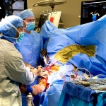
For patients interested in optimal placement of their hip and knee arthroplasty components, the orthopedic community has used the Mako robotic-assisted surgery system for many years. This technology has allowed hip and knee reconstruction surgeons to very accurately carry out total knee arthroplasty and total hip arthroplasty. Over the past year, the Mako system has been introduced to reverse shoulder arthroplasty surgery. But what exactly is Mako robotic-assisted reverse shoulder arthroplasty?
A Mako RSA is the exact same surgery as a traditional RSA; however, the technique used to prepare the glenoid is performed using a robotic arm. This makes execution of the pre-surgical plan very accurate—not only planning out the surgery in 3D ahead of time, but also carrying out that exact plan unique to each patient.
The main differences between a Mako RSA and a traditional RSA are:
1. Placement of a checkpoint device.
The checkpoint is impacted into the superior glenoid or base of the coracoid bone to act as a reference during surgery. I now prefer the superior glenoid placement because that location has dense bone and is very secure.
2. Placement of a coracoid array.
The coracoid array is clamped to the coracoid process of the scapula bone. This must be very secure or the Mako system will not “know” where the socket is located. The Mako visualization camera “sees” where the scapula bone is located in the operating room by referencing the markers on the array. I like to tighten the clamp manually to make sure it is very stable, but not over-tightened.
3. Registration of the bony socket.
The bone the glenoid (socket) is “registered” or connected to the Mako system using a probe with markers that can be visualized by the camera. The registration process links the patient’s actual glenoid, or socket, to the computer system.
4. Bony preparation: the robotic arm then uses a high-speed bur to prepare the glenoid bone. The robotic arm has a haptic feedback system to let me know when I’m at the limits of the surgical plan. If the bur is not at the proper location on the socket, the Mako system will simply stop rotating to protect areas outside of the planned reconstruction. The Mako robotic arm acts as a guardrail to ensure that only the proper amount of bone is removed to execute the plan accurately and completely. This includes preparation of the glenoid bone, central boss, and central fixation with a screw or post.
5. I make the deltopectoral incision approximately 1 cm more proximal to allow for adequate exposure of the coracoid.
6. I perform extensive pericoracoid releases to allow for optimal registration and coracoid clamp array placement.
7. I perform a partial coracoacromial ligament release, as minimally as possible, to facilitate optimal coracoid array clamp placement and security.
8. I now prepare the central fixation, typically with a screw, before preparing the glenoid face and central boss. This helps ensure that the overall plan is properly centered on the glenoid face.
Everything else for a Mako reverse replacement is very similar to a traditional reverse shoulder replacement. I use a deltopectoral approach for both traditional and robotic-assisted RSA surgeries. The long head of the biceps tendon is managed according to the patient’s needs. Extensive periglenoid releases are performed. The subscapularis is mobilized, releases are performed as needed, and repair is performed if possible. Bone contouring is performed with tuberoplasty for the lesser and/or the greater tuberosity if impingement is identified during trialing. The shoulder is tested to optimize impingement-free motion, stability, and proper positioning.
In my opinion, the potential benefits of reverse shoulder arthroplasty using Mako robotic-assisted technology primarily involve avoiding outliers. As in any highly precise endeavor, outliers are a problem. If we think of a sport like basketball, the best free-throw shooters in the world shoot over 90% from the free-throw line, but very few people actually make 100% of their shots—especially during a high-stakes professional game. In medicine, however, even a small mistake can be catastrophic for a patient. Outliers must be avoided whenever possible. I always aim for 100% accuracy.
Numerous checks and balances are used during surgery to help avoid outliers. Mako robotic-assisted RSA is a strong tool to help ensure accurate placement of the 3D preoperative surgical plan that I perform in Blueprint. There are other ways to achieve very accurate surgery. These include the use of 3D preoperative planning, patient-specific instrumentation, robot-assisted surgery, and patient-specific implants that are 3D printed. In my current practice, Mako robotic-assisted RSA surgery plays a significant role in helping me achieve very accurate surgery and getting as close as possible to 100% for my patients.
I perform these surgeries regularly at the Orlando Health Jewett Orthopedic Institute. Please reach out to info@benservicemd.com with any questions regarding Mako robotic-assisted RSA surgery.